在上篇中,我们介绍了人体处于站立位时,EOS系统、颅骨的影像学标记、颈椎角度和胸腰椎角度的测量方法。在下篇中,我们将通过对骨盆环角度、下肢角度、前方失平衡的代偿机制的分析,全面讨论从头到足的脊柱平衡状态,同时通过运动学分析将人体行走时的脊柱动态状况呈现出来,有助于判断患者脊柱的真实状态。
(五)骨盆环的角度
Jean Dubousset等认为骨盆环是脊柱和下肢之间的一个中间椎体。
Duval-Beaupère[3]在侧位X线上描述3个骨盆的角度。
● PI角(骨盆入射角),由股骨头中点与S1终板中点的连线和一条经过S1椎体上终板中点并垂直于S1终板的线所构成的夹角。这个角度描述了骨盆在前后方向的形状和宽度,它决定了脊柱的弯曲,尤其是腰椎前凸。PI在生长过程中逐渐增加[22]且由遗传因素决定。它不随骨盆的位置而变化,理论上也不随年龄而变化。事实上,最近的研究表明[23]PI可以随着年龄的增长而增加,这是由于位于股骨头(或髋臼)和骶骨之间的骶髂关节松弛造成的。
● PT角(骨盆倾斜角),是由经过股骨头中点的垂线和连接股骨头中点与S1椎体上终板中点的直线所构成的夹角。当骨盆后倾时,PT角增加,因为这是对脊柱前方失平衡的一种自动纠正,在骨盆前倾时,PT角度减小。
● T9矢状位悬垂角,由经过股骨头中心的垂线与股骨头和T9椎体中点的连线所构成的夹角。Duval-Beaupère认为T9椎体中点是人体躯干的重心。这个角度定位躯干,而不是骨盆,PT角决定了骨盆的位置。T9矢状位悬垂角在很长一段时间保持不变,当出现严重前方失平衡时,此角度会减小甚至负值。
(六)下肢角度
Itoi[24]补充了一些关于骨质疏松症患者的代偿角度的概念——股骨胫骨角,由股骨干轴线和胫骨干轴线相交构成。当膝盖弯曲的时候,该角是正值,这是脊柱代偿严重向前失平衡的第二种方式。
Mangione和Sénégas[25]使用股骨骨盆角来评估重度前方失平衡时髋关节的伸展程度,它由股骨干轴线和股骨头中心与骶骨终板中点的连线所构成。
Hovorka等[26]和Lazennec等[27]通过SlotX线片研究髋关节伸展以评估髋关节的功能,其在前方严重失平衡的情况下能够代替骨盆后倾。
(七)前方失平衡的代偿机制
本章节讨论了从头到足的脊柱平衡状态,C7铅垂线对于平衡的判定时常不准确,因为这种方式没有考虑到头部对于整体平衡的影响。把颅骨和骨盆看作是一个整体对于研究平衡状态至关重要。
因此,我们可以通过评估外耳道(头骨的重心)和股骨头(股骨头中心,几乎是骨盆的中心)的连线来实现。Gangnet等[28]证明了这两个点(外耳道和股骨头)在正常骨盆形态和处于伸膝状态(完美平衡)的健康受试者中是垂直对齐的。
而对于通过骨盆后倾代偿平衡的患者,股骨头中点会移动到外耳道点的前下方,随后膝关节屈曲,躯干后移,最终胸椎后凸角度减小,有时还会出现上位腰椎向后滑移。PI角大的人群骨盆后倾的能力更强。
Sénégas等[15]讨论了使用踝关节过伸的方式来纠正前方失平衡的方法。尽管有时整个过程对患者来说往往是难以忍受的,但是当从外耳道引下的垂直轴自然落在股骨头前面时,我们把这称为前方失平衡。
外耳道-股骨头垂直对齐可能在某些疾病中持续很长时间,这使得作者定义了向上或向下对线,可用于保持活动度的脊柱节段,主要是颈椎。
向上力线解释了合并颈椎后凸的特发性脊柱侧弯患者,通常合并有平背表现(图3-36)。
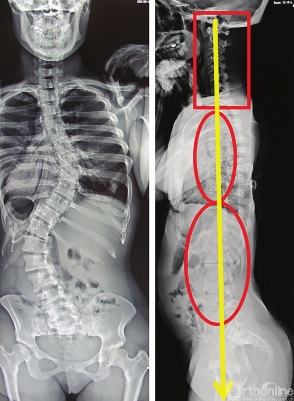
图3-36 特发性脊柱侧弯“平背”患者的脊柱力线,颈椎呈后凸状态
向上力线的其他表现包括矫正腰椎后凸的患者接受大型截骨术矫正后,颈椎前凸角度增加(图3-37,图3-38)。
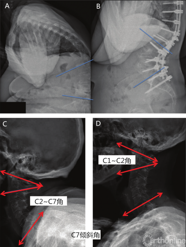
图3-37 腰椎截骨术后的“向上力线”
A.术前腰椎X线;B.术后腰椎X线;C.术前颈椎X线;D.术后颈椎X线。C2~C7角减小较多,C1~C2角减小较小
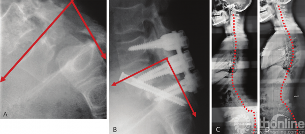
图3-38 发育不良性脊柱滑脱术后的脊柱力线
A.术前腰骶椎X线;B.术后腰骶椎X线;C.全脊柱术前X线;D.全脊柱术后X线
手术治疗scheuermann病重建胸椎后凸导致颈椎前凸角度减小,腰骶后凸并伴有发育不良性脊柱滑脱者会出现腰前凸角度减小和胸椎后凸的增加(图3-39,图3-40)。
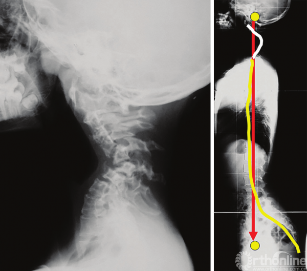
图3-39 神经纤维瘤病患者伴有颈椎严重后凸的脊柱力线,可以注意到胸椎变平以获得外耳道与股骨头之间力线的最佳匹配
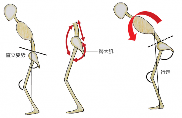
图3-40 行走时,由于臀大肌无法维持骨盆后倾,矢状位失平衡加剧
向下对线很少见,在神经纤维瘤病伴严重的颈椎后凸畸形,可观察到较少见的向下对线,导致胸椎平背畸形(图3-41)。
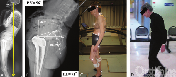
图3-41 A.静态后方失平衡患者;B.PI角大,后倾代偿幅度大的患者;C.步态分析;D.患者行走过程中伴有严重的矢状位失平衡
行走评估
整个脊柱的静态X线片并不代表运动过程中的真实状态。Lee等[29]研究表明,与静态侧位图像相比,术后平背患者行走时的前倾失平衡更为明显。一个可能的解释是,由于臀大肌肌肉力量减弱,在行走时不能够维持骨盆极度后倾状态。代偿现象在静态研究中起到了很大的作用,但并不能在动态研究中起到作用[30]。
有些患者存在过度后倾失平衡,表现为他们觉得“坐在自己的骨盆上”,外耳道铅垂线落在股骨头后方,行走时躯干向前倾斜。腰椎截骨术可以极大改善这些患者的状态(图3-40)。
人体行走时,对脊柱进行动态分析是非常复杂的。它需要通过运动学分析来测量各部分在空间中的位置,该运动学分析需要与对躯干肌(竖脊肌和腹肌)和臀肌(主要是臀大肌)的活动分析相结合。
实验人员通过在受试者骨骼表面的对应位置放置传感器(设置标记点)来实施运动分析。这些位置由一系列红外摄像机永久记录,并通过专用的软件进行分析,在三维空间上(x、y、z)精确的描述脊柱的位置。除了运动的幅度,研究者还可以决定一个步行周期,从足跟接触开始,以对侧足跟接触结束。因此也可以确定步态参数,如步速、宽度、长度、频率的规律性。
研究人员可以通过表面肌电图对肌肉进行分析。传感器与运动分析传感器耦合,以促进受试者运动和肌肉活动之间的联系。
矫形手术对于运动参数的改善有很大帮助,其与脊柱矢状面平衡的恢复呈正相关[31]。然而,手术后患者的步态与健康受试者仍存在差异。因矢状面失平衡而接受翻修手术的患者中,步行功能的恶化更为严重[32]。术后步态参数是评估干预效果的客观指标,是对上述影像学评估的重要补充[33]。(完)
参考文献
[1] Luboga SA, Wood BA. Positionand orientation of the foramen magnum in higher primates. Am J Phys Anthropol.1990; 81(1):67–76
[2] Tardieu C, Bonneau N, HecquetJ, et al. How is sagittal balance acquired during bipedal gait acquisition?Comparison of neonatal and adult pelves in three dimensions.
Evolutionary implications. J HumEvol. 2013; 65(2): 209–222
[3] Duval-Beaupère G, Schmidt C,Cosson P. A barycentremetric study of the sagittal shape of spine and pelvis:the conditions required for an economic standing position. Ann Biomed Eng.1992; 20(4):451–462
[4] Morvan G, Wybier M, Mathieu P,Vuillemin V, Guerini H. Clichés simples du rachis: statique et relations entrerachis et bassin [in French]. J Radiol. 2008; 89(5 Pt 2):654– 663, quiz 664–666
[5] Schlösser TPC, Janssen MMA,Vrtovec T, et al. Evolution of the ischio-iliac lordosis during natural growthand its relation with the pelvic incidence. Eur Spine J. 2014; 23(7):1433–1441
[6] Abitbol MM. Evolution of thelumbosacral angle. Am J Phys Anthropol. 1987; 72(3):361–372
[7] Marty C, Boisaubert B, DescampsH, et al. The sagittal anatomy of the sacrum among young adults, infants, andspondylolisthesis patients. Eur Spine J. 2002; 11(2):119–125
[8] Goff CW, Landmesser W. Bipedalrats and mice; laboratory animals for orthopaedic
research. J Bone Joint Surg Am.1957; 39-A(3):616–6–22
[9] Aylott CEW, Puna R, RobertsonPA, Walker C. Spinous process morphology: the effect of ageing throughadulthood on spinous process size and relationship to sagittal alignment. EurSpine J. 2012; 21(5):1007–1012
[10] Hadar H, Gadoth N, Heifetz M.Fatty replacement of lower paraspinal muscles: normal and neuromusculardisorders. AJR Am J Roentgenol. 1983; 141(5):895–898
[11] Cruz-Jentoft AJ, Baeyens JP,Bauer JM, et al. Sarcopenia: European consensus on definition and diagnosis:report of the European Working Group on Sarcopenia in Older People. Age Ageing.2010; 39(4):412–423
[12] Fortin M, Videman T, GibbonsLE, Battié MC. Paraspinal muscle morphology and composition: a 15-yrlongitudinal magnetic resonance imaging study. Med Sci Sports Exerc. 2014;46(5):893–901
[13] Vital JM, Gille O, Coquet M.Déformations rachidiennes: anatomopathologie et histoenzymologie [in French].Rev Rhum. 2004; 71:263–264
[14] Sénégas J, Bouloussa H,Liguoro D, Yoshida G, Vital JM. Evolution morphologique et fonctionnelle durachis vieillissant. In: Anatomie de la Colonne Vertébrale: Nouveaux Concepts(in French). Montpellier, France: Sauramps Médical; 2016:111–155
[15] Solow B, Tallgren A. Naturalhead position in standing subjects. Acta Odontol Scand. 1971; 29(5):591–607
[16] Peng L, Cooke MS. Fifteen-yearreproducibility of natural head posture: A longitudinal study. Am J OrthodDentofacial Orthop. 1999; 116(1):82–85
[17] Sugrue PA, McClendon J, Jr,Smith TR, et al. Redefining global spinal balance: normative values of cranialcenter of mass from a prospective cohort of asymptomatic individuals. Spine.2013; 38(6):484–489
[18] Vital JM, Sénégas J.Anatomical bases of the study of the constraints to which the cervical spine issubject in the sagittal plane. A study of the center of gravity of the head.Surg Radiol Anat. 1986; 8(3):169–173
[19] Vidal J, Marnay T. Sagittaldeviations of the spine, and trial of classification as a function of thepelvic balance (in French). Rev Chir Orthop Repar Appar Mot. 1984; 70 Suppl2:124–126
[20] Delmas A, Depreux R. Spinalcurves and intervertebral foramina (in French). Rev Rhum Mal Osteoartic. 1953;20(1):25–29
[21] Roussouly P, Gollogly S, BerthonnaudE, Dimnet J. Classification of the normal variation in the sagittal alignmentof the human lumbar spine and pelvis in the standing position. Spine. 2005;30(3):346–353
[22] Mangione P, Gomez D, SénégasJ. Study of the course of the incidence angle during growth. Eur Spine J. 1997;6(3):163–167
[23] Jean L. Influence of age andsagittal balance of the spine on the value of the pelvic
incidence. Eur Spine J. 2014;23(7):1394–1399
[24] Itoi E. Roentgenographicanalysis of posture in spinal osteoporotics. Spine. 1991; 16(7):750–756
[25] Mangione P, Sénégas J.Sagittal balance of the spine (in French). Rev Chir Orthop Repar Appar Mot.1997; 83(1):22–32
[26] Hovorka I, Rousseau P,Bronsard N, et al. Extension reserve of the hip in relation to the spine: Comparativestudy of two radiographic methods (in French). Rev Chir Orthop Repar Appar Mot.2008; 94(8):771–776
[27] Lazennec JY, Charlot N, GorinM, et al. Hip–spine relationship: a radio-anatomical study for optimization inacetabular cup positioning. Surg Radiol Anat. 2004; 26(2):136–144
[28] Gangnet N, Pomero V, Dumas R,Skalli W, Vital JM. Variability of the spine and pelvis location with respectto the gravity line: a three-dimensional stereoradiographic study using a forceplatform. Surg Radiol Anat. 2003; 25(5)(–) (6):424–433
[29] Lee CS, Lee CK, Kim YT, HongYM, Yoo JH. Dynamic sagittal imbalance of the spine in degenerative flat back:significance of pelvic tilt in surgical treatment. Spine. 2001;26(18):2029–2035
[30] Shiba Y, Taneichi H, Inami S, MoridairaH, Takeuchi D, Nohara Y. Dynamic global sagittal alignment evaluated bythree-dimensional gait analysis in patients with degenerative lumbarkyphoscoliosis. Eur Spine J. 2016; 25 (8):2572–2579
[31] Yagi M, Kaneko S, Yato Y,Asazuma T, Machida M. Walking sagittal balance
correction by pedicle subtractionosteotomy in adults with fixed sagittal imbalance.
Eur Spine J. 2016; 25(8):2488–2496
[32] Engsberg JR, Bridwell KH,Reitenbach AK, et al. Preoperative gait comparisons between adults undergoinglong spinal deformity fusion surgery (thoracic to L4, L5, or sacrum) andcontrols. Spine. 2001; 26(18):2020–2028
[33] Engsberg JR, Bridwell KH,Wagner JM, Uhrich ML, Blanke K, Lenke LG. Gait changes as the result ofdeformity reconstruction surgery in a group of adults with lumbar scoliosis.Spine. 2003; 28(16):1836–1843, discussion 1844
来源:《脊柱矢状位平衡》
翻译:吴 兵 于 洋
校对:钱邦平 吕国华
主译:海 涌 李 利 李危石 郑召民
主审:邱 勇 吕国华
点击下图查看书籍简介↓
声明:此文内容及图片由供稿单位提供,仅供学习交流,不代表骨科在线观点。
相关阅读: